Oral Roentgenology : a Roentgen study of the anatomy and pathology of the oral cavity (1917)
Thoma, Kurt H. (Kurt Hermann), 1883- · Boston, Mass. : Ritter & Company · leaf 74
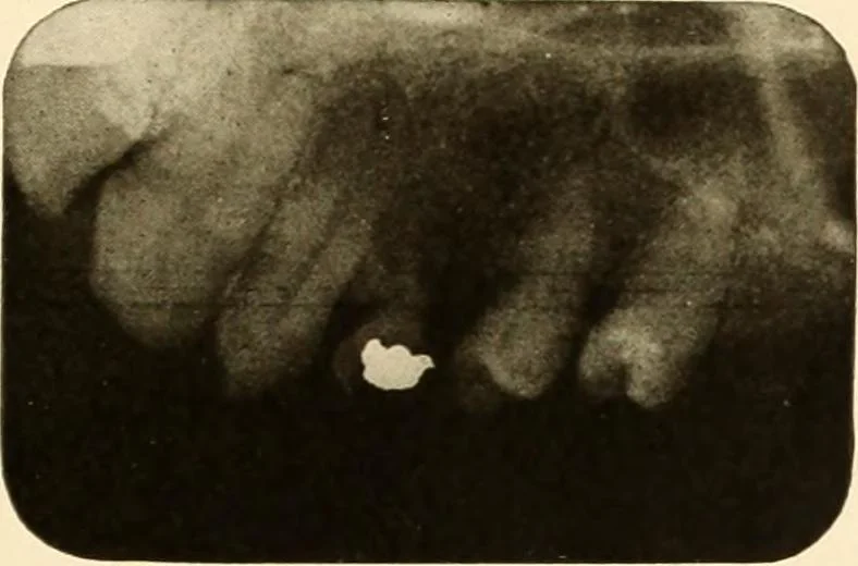
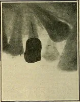
mporarymolars have been retained. The same is true of the left lower side.In neither of the pictures is there any evidence of the permanent teeth.Also, on the right lower side, the bicuspids are absent. The temporaryteeth have been shed, except for a small piece of root. Figure 67. Patient: Miss B.… … Figure 62.